
Apoptosis is an active, programmed process of autonomous cellular dismantling that avoids eliciting inflammation. Necrosis, on the other hand, is characterized as passive, accidental cell death resulting from environmental perturbations, resulting in the uncontrolled release of inflammatory cellular contents. During apoptosis, calcium-dependent phosphatidylserine (PS) is transferred to the outer leaflet of the plasma membrane. As a universal indicator of the early and intermediate stages of apoptosis, the appearance of PS on the cell surface can be detected before morphological changes are observed.
Annexin V Staining
Researchers in many fields often want to know whether apoptosis is happening or not, while those dedicated to studying apoptosis usually also want to decipher when/under what conditions it is triggered or prevented, and what is triggering or preventing it.
The most straightforward way of detecting apoptosis is through the use of Annexin V-specific stains, which can detect PS on the outer surface of the plasma membrane during early and intermediate apoptosis. The Annexin V approach is easy, fast and cost-effective, making it the most widely-used apoptosis assay today.
Pulling Apart The Stages Of Apoptosis with Multiplex Detection Kits
However, while Annexin V staining provides useful information about the occurrence of apoptosis, it alone doesn’t allow one to discriminate between healthy, dying and dead cells, which is a drawback for those who want delineate the events and mechanisms underpinning apoptosis in more detail.
AAT Bioquest’s Cell Meter™ Multiplexing Live, Apoptotic and Necrotic Cell Detection Kit IIIemploys a three-probe system to simultaneously monitor apoptotic, necrotic and healthy cells with a fluorescence microscope.
How Does The Multiplex Kit Work?
Alive Cells
Viable cells are detected and labeled using AAT Bioquest’s cell-permeant dye Cellbrite™ Red (Ex/Em = 613/631nm).
Dying Cells
Cells undergoing the early and intermediate stages of apoptosis are detected and labeled with PS sensor Annexin V-iFluor™ 488 (Ex/Em = 494/520 nm). Upon association with membrane-exposed PS, Annexin V-iFluor™ 488 will fluoresce green.
Dead Cells
Loss of plasma membrane integrity, as demonstrated by the ability of a membrane-impermeable Nuclear Blue™ DCS1 (Ex/Em = 350/461 nm) to label the nucleus, reveals cells in the late stage of apoptosis and necrosis in a straightforward way.
The fluorescence signal generated by each of these three probes is proportional to the number of apoptotic (green), necrotic (blue) or viable (red) cells, respectively.
What Can You Expect? Sample Data
Figure 1 below displays fluorescence images of HeLa cells labeled with the Cell Meter™ Multiplexing Kit. Setup: HeLa cells at a density of 100,000 cells/well/100 µL were seeded overnight in a 96-well black wall/clear bottom plate.
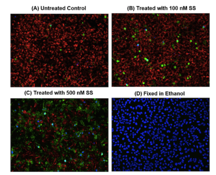
Figure 1. Cells were treated with 0-500 nM staurosporine* (SS) at 37ºC for 4 hours (A-C), or fixed in ethanol (D), then incubated with the triple fluorescence assay solution for 1 hour. The fluorescence signal was measured using a fluorescence microscope with a Cy5 filter for healthy cells (Red), FITC filter for apoptotic (Green) and DAPI filter for necrotic cells (Blue), respectively. * Staurosporine is a broad-spectrum kinase inhibitor that is widely used as a positive control in studies of apoptosis.
Want to Know More?
For more information on this kit, or any other tools to study apoptosis and necrosis, feel free to get in touch with our team at info@nordicbiosite.com.